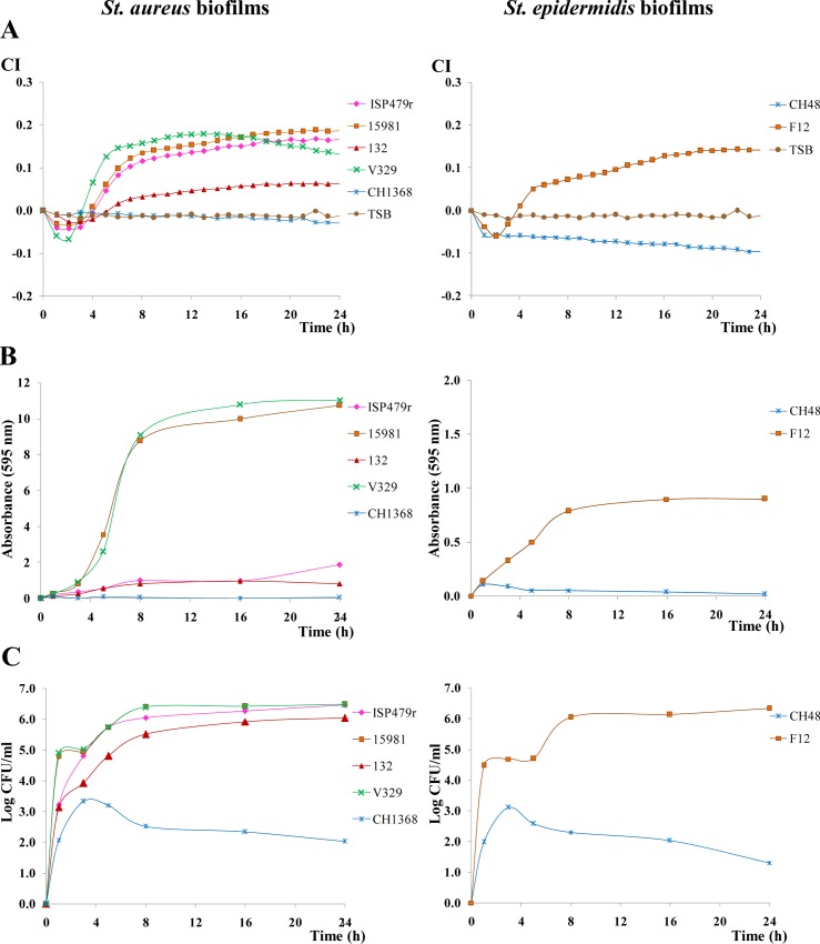
Fig 4. Variation in the cell index (CI) during biofilm formation at 37°C of different S. aureus and S. epidermidis biofilm producers (ISP479r, 15981, 132 or V320 and F12, respectively) and no-biofilm producers (CH1368 and CH48, respectively).
TSB+0.25% glucose was the culture medium used in the experiment (A). Absorbance (595 nm) measured after crystal violet staining of samples collected at different times during the biofilm formation in E-plates of the strains under study (B). Counts (Log CFU/ml) of cells collected from the biofilms formed in the E-plates by the strains under study (C). Statistical differences among strains at three sampling points (8, 16 and 24 h) are collected in S1 Table, which also shows representative mean and SD values.

