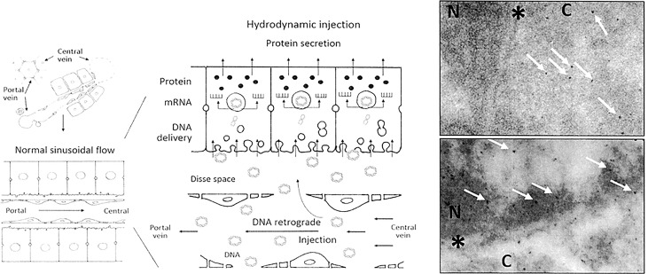
Fig 6. Histological and cellular scheme of hydrofection and nanoparticles distribution within liver tissue.
The schematic area (left) provides a representation of how hydrodynamic retrograde injection, under the described conditions, mediates greater separation between endothelial cells and their fenestrations, as well as enlargement of the Disse space and the formation of massive endocytic vesicles. These changes result in DNA molecule entry into the liver cell nucleus, where DNA transcription to RNA occurs. Then, mRNA is released into the cell cytoplasm, where it is translated to protein. Finally, after the necessary modifications, the mature protein, if to be secreted, is released out of the tissue. The image at right provides a transmission electron microscopy view of the nuclear compartment of hepatocytes after transferring 4 nm and 15 nm gold nanoparticles by the surgery closed procedure (lower panel) and open procedure (upper panel). Nanoparticles only reach the nucleus with more energetic conditions of the open procedure. N: nucleus; C: cytoplasm; *: nuclear envelope. White arrows: nanoparticles 4–8 nm in diameter.

