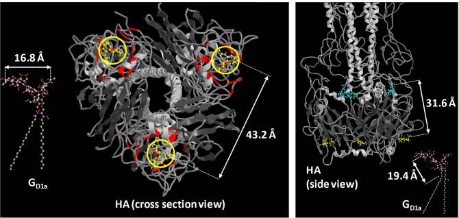
Fig 7. Using structural arguments to understand binding results.
Left) View of HA protein head group in relation to GD1a. Red regions show the binding pockets and yellow circles show where sialic acid are located. Right) Side view of HA protein in relation to GD1a. Teal molecules are sialic acids at potential secondary binding sites. The SLB would be on the bottom side of the protein, while the viral membrane would be on the top side. The hemagglutinin structure and sialic acid positions were obtained by Sauter et al. [65] PDB ID: 1HGG.

