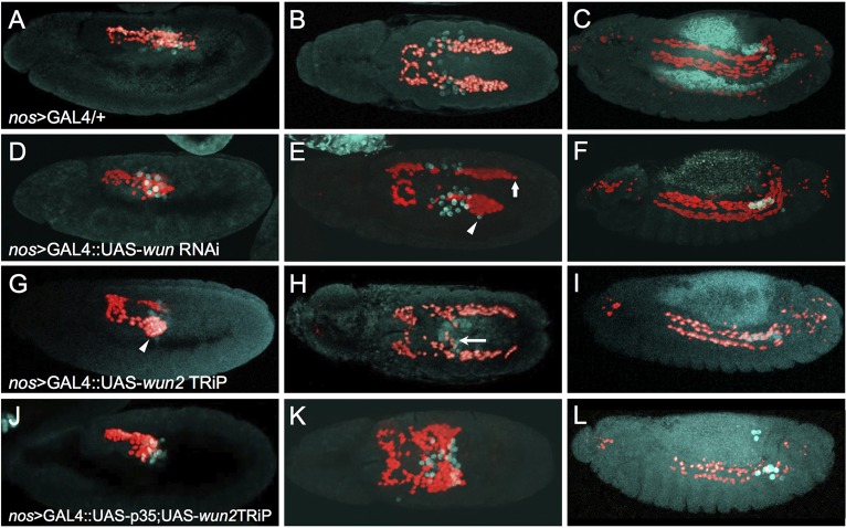
Fig. 3.
Germ cell-derived Wun is required for proper CVM cell migration. Immunofluorescence on embryos expressing HC3 to detect CVM (anti-RFP; red) and PGCs (anti-Vasa; cyan) in early stage 11 (A,D,G,J, lateral view), late stage 11 (B,E,H,K, dorsal view) and stage 13 (C,F,I,L, lateral view). All embryos express nos>GAL4.VP16 (nos>GAL4), which activates expression of UAS constructs specifically in the PGCs at these stages of development. (A-C) The nos>GAL4.VP16 driver alone shows wild-type CVM cell and PGC migration. (D-F) UAS-wun6446 RNAi driven by nos>GAL4.VP16 shows a weak phenotype, if any, as asymmetric migration (arrow) and a few lost PGCs (arrowhead) were observed at late stage 11. (G-I) Knocking down wun2 expression in PGCs by expression of UAS-wun232423TRiP causes the CVM cells to aggregate on the lateral side of the pmg in early stage 11 (arrowhead, G) and to mismigrate towards PGCs at late stage 11 (H, arrow), as well as causing a loss of CVM cells and PGCs at stage 13 (I). (J-L) Coexpressing the anti-apoptotic factor p35 along with the wun232423TRiP causes severe mismigration phenotypes. PGCs exit the pmg (J) but subsequently stall (K), resulting in sequestration of CVM cells at this position (K). More PGCs remain alive upon coexpression of p35 (compare L and I; see Fig. S4) but cause the opposite effect on CVM cells, as fewer cells are present at stage 13 (L). Presumably, more CVM cells die because they fail to reach the appropriate position along the TVM or otherwise mismigrate and die.

