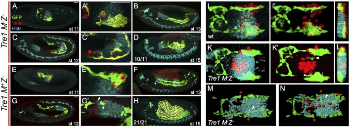
Fig. 4.
Immobilization of germ cells within the pmg redirects CVM cell migration. Lateral view (A-H) or dorsal view (I-N) of embryos fluorescently stained for GFP (GV2, CVM), Vasa (PGCs) and Hb9 (midgut primordium) in Tre1 mutants that are either M−Z− (A-D,K-N) or M+Z− (E-H) or wt (GV2/+) (I,J). Scale bars in A′,C′,E′,G′ show relative magnification to images in A,C,E and G, respectively. (A,E) Stage 11 embryos show colocalization of PGCs and CVM internal (A,A′) or just external (E,E′) to the Hb9 (pmg-associated endoderm) staining. (C,G) In stage 12 Tre1 M−Z− mutants (C,C″) most if not all of the PGCs are still internal to the pmg and the CVM migration does not continue past the end of the midgut, whereas in the M+Z− Tre1 mutants (G,G′) there are a few PGCs trapped in the pmg (arrowhead) and CVM cells are associated with them (arrow), although the majority of the CVM cells migrate normally. (B,F) At stage 13, the CVM in the Tre1 M−Z−mutants (B) fails to migrate to the anterior end of the pmg (marked by arrowheads in B and F) and all but one PGC remain internal to the pmg, whereas in the M+Z− mutants the PGCs have populated the gonadal mesoderm and the CVM appears somewhat normal except for cell death in the posterior (arrow). (D,H) The CVM in stage 15 Tre1 M−Z− mutants (D) fails to cover the gut and most if not all of the PGCs are lost. Ten out of the eleven embryos examined at this stage showed this severe phenotype, whereas 21 out of 21 M+Z− Tre1 mutant embryos examined look grossly normal at stage 15 (H). (I-L) Stage 11 embryos showing GV2/+ (I,I′) or Tre1 M−Z− (K,K′). (J,L) yz slice through the center of the pmg, with dorsal to the right, shows that the CVM in GV2/+ embryos migrates over the dorsal side of the pmg (I,J), whereas in the Tre1 M−Z− mutants the CVM cells invade the center of the pmg where the PGCs are localized (K,L, arrowheads in K′). (M,N) 3D projection model of stage 12 Tre1 M−Z− embryo created in Imaris using the surface detector (for CVM and pmg) or the spot detector (for PGCs) to clearly delineate the three cell groups. Arrowhead indicates CVM cells that are internal to the pmg when viewed in a single embryo both from the dorsal (M) and ventral (N) side of the projection.

