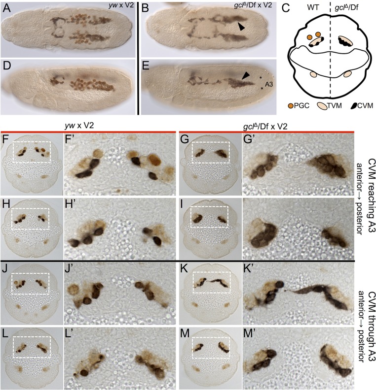
Fig. 5.
CVM cells are more tightly organized in the absence of germ cells. (A,B,D,E) Immunostaining for CVM (anti-GFP, black) and germ cells (anti-Vasa, brown) in embryos from wild type (yw) females (A,D) and from gclΔ/Df maternal mutants (B,E) crossed to GV2 males. (A,B) Dorsal views; (D,E) lateral views. Arrowheads (B,E) indicate regions of tighter CVM association. (C) Schematic depicting the spatial relationship between CVM, TVM and germ cells in a transverse section of a stage 11 embryo, near the back of the CVM cluster that overlaps the germ cells on the anterior-posterior axis. An embryo from a wild-type female is depicted on the left half of the section, and an embryo from a germ cell-less embryo from a gclΔ/Df maternal mutant on the right half of the section. (F-M′) Transverse sections of embryos immunostained for CVM (black, anti-GFP), PGCs (anti-Vasa, strong brown) and TVM (anti-FasIII, pale brown) of the indicated genotypes. F′-M′ show a magnified view of the boxed regions. CVM is more widely spaced in embryos from yw females (F,H,J,L) as compared with the CVM that is more tightly clumped and forms multilayers in embryos from gclΔ/Df females (G,I,K,M). Staging of embryos was matched by determining the progress of CVM cells through the abdominal segments. Asterisks (E) mark the approximate boundaries of the third abdominal segment (A3) for reference of CVM progression in F-M.

