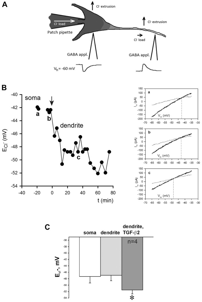
Fig. 4.
Treatment with TGF-β2 augments extrusion of intracellular Cl− from cultured hippocampal neurons. (A) Schematic representation of the experimental paradigm for assessing changes in dendritic chloride extrusion. GABA is locally applied at the soma and at a primary dendrite (100 µm distal from the soma) of a neuron recorded in whole-cell voltage clamp mode with a pipette containing a slight load of chloride. The reversal potential in for GABA is estimated in the two positions. (B) Timecourse of Cl− reversal potential measured in soma (a) and dendrite (b,c) of a cultured (DIV 10) hippocampal neuron. Onset of TGF-β2 application corresponds to time point zero. In this particular neuron, almost no somatodendritic Cl− gradient was observed before exposure to TGF-β2 as the difference in ECl− measured in soma (a) and in dendrite (b) was almost zero. After application of TGF-β2, dendritic ECl− and therefore somatodendritic Cl− gradient became negative (c). The insets are example traces of voltage ramps before (light gray) and during local application of GABA (dark gray) at the soma (a) and the dendrites (b,c). The intercept between these traces gives an estimation of the GABAA reversal potential at the specific location. b and a are example traces before and after application of TGF-β. (C) Quantification of dendritic ECl− in control and TGF-β2-treated neurons. *P<0.05 compared to the control (unpaired Student's t-test). Error bars represent s.e.m. from four independent experiments. Data used in this panel include only those experiments where no statistically significant difference between somatic and dendritic ECl− measured in the same cell was observed prior to TGF-β2 application (Fig. S1).

