Fig. 5.
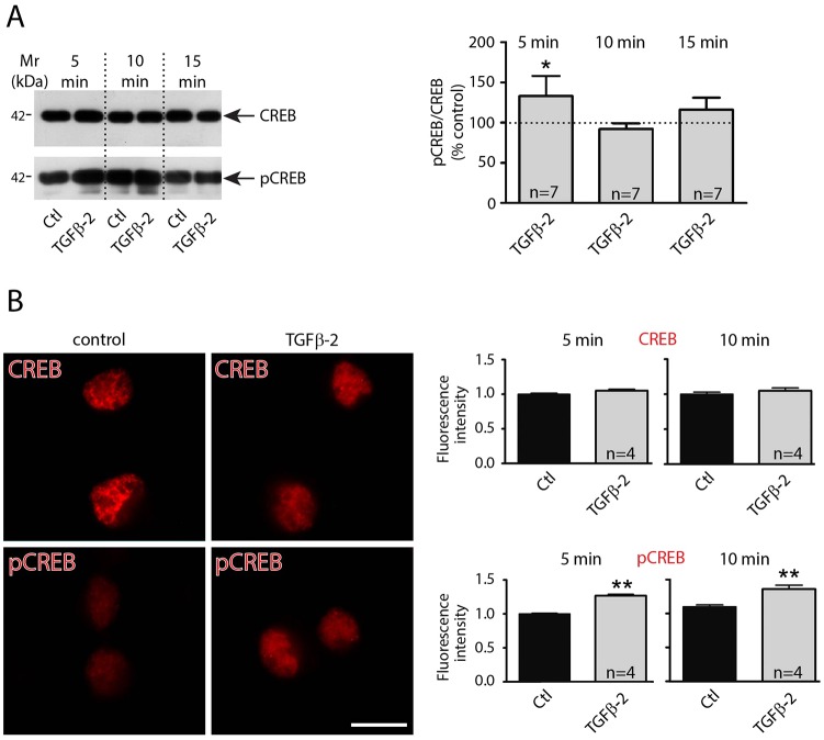
TGF-β2 activates CREB by increasing its phosphorylation. (A) Western blot analysis of total and of phosphorylated CREB in cultured hippocampal neurons (DIV12) treated with 2 ng/ml TGF-β2 for either 5, 10 or 15 min (dotted line represents values for control). (B) Immunofluorescence for CREB1 and pCREB of mouse hippocampal cultures (DIV12) under control conditions and following treatment with TGF-β2 for 10 min. Scale bar: 10 µm. Quantification of relative CREB1 and phospho-CREB fluorescence intensity following application of TGF-β2 for 5 and 10 min (images are representative out of four experiments). Data are shown as mean±s.e.m. for seven or four experiments as indicated. *P<0.05, **P<0.01 (unpaired Student's t-test).

