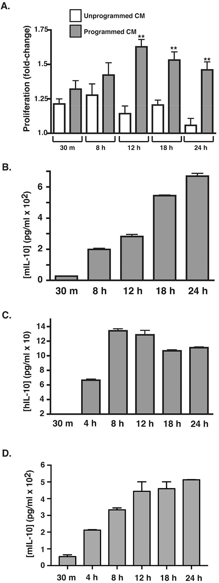
Fig. 2.

Timing of induction of the pathological macrophage phenotype by ADPKD programming coincides with induction of IL-10 secretion. (A) RAW macrophages were incubated in duplicate with medium or ADPKD-CM for the indicated times. Media were collected and stored at 4°C, and cells were then washed and incubated with basal medium for 24 h to produce programmed or unprogrammed CM, which were then assayed for the ability to stimulate proliferation of PKD-A cells, as described in Materials and Methods. Data are presented as the fold change relative to the baseline proliferation determined from PKD-A cells grown in 0.1% FBS in parallel. Similar results were observed in three independent experiments using CM generated with cyst cells from different ADPKD individuals. (B) RAW-macrophage-produced IL-10 levels were measured in the collected ADPKD-CM that was used for the programming described in A with the use of an ELISA kit specific for mouse IL-10. Similar results were observed in three independent experiments using CM generated with cyst cells from different ADPKD individuals. (C) THP-1 macrophages were incubated with ADPKD-CM for the times indicated, and IL-10 secretion was measured by using ELISA. Human IL-10 was not detectable in either the ADPKD-CM used for programming or in medium from unprogrammed macrophages (data not shown). (D) Mouse BMDMs were programmed in duplicate for the time indicated with primary NHK-conditioned medium. Media were collected, and IL-10 concentration was measured by using ELISA. hIL-10, human IL-10; mIL-10, mouse IL-10. Data are mean±s.e.m. **P<0.01 (two-tailed t-test).
