Fig. 5.
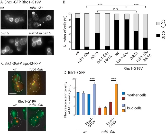
Constitutively active Rho1 restores GFP–Snc1 transport and Bik1 association to microtubule plus-ends. (A) Effect of Rho1-G19V expression on GFP–Snc1 localization in different strains as indicated. Rho1-G19V rescue of Snc1 misrouting is dependent on the presence of Bik1. (B) Quantification of the localization of GFP–Snc1 pattern either at the bud plasma membrane and in cytoplasmic dots, or at mother and bud plasma membranes in the different strains (wt, n=60; tub1-Glu, n=51; bik1Δ, n=59; tub1-Glu bik1Δ, n=45; wt-Rho1-G19V, n=55; tub1-Glu-Rho1-G19V, n=131; bik1Δ- Rho1-G19V, n=144; tub1-Glu bik1Δ Rho1-G19V, n=183 cells). ***P<0.0001; n.s., not significant (Fisher's exact test). (C) Distribution of Bik1–3GFP in wt and tub1-Glu cells expressing Rho1-G19V. The spindle pole body was labeled by co-expression of the Spc42–RFP protein. (D) Quantification of Bik1 fluorescence intensity at microtubule plus-ends in mother or bud cells (without and with Rho1-G19V, respectively: wt, n=49 and 69 cells, tub1-Glu, n=56 and 78 cells). Results are mean±s.e.m. ***P<0.0001 (two-tailed Mann and Whitney test). Scale bars: 5 µm.

