Fig. 5.

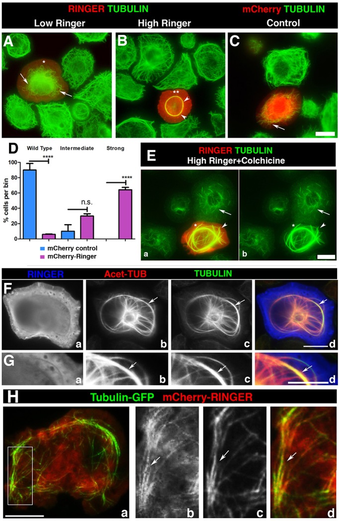
Ringer affects the morphology and stability of microtubules in cells. (A–C) mCherry-tagged Ringer (Ringer) and control mCherry alone (mCherry) was expressed in S2 cells, which were immunostained for Tubulin. Low Ringer expression (asterisk) leads to abnormal microtubule curvature and failure to grow into cell periphery (A, arrow). (B) At high levels of Ringer expression (asterisks), microtubules were incorporated into a central ring-like bundle (arrowheads) not seen in mCherry controls (C, arrow). (D) Quantification of microtubule phenotypes (n≥100). Individual cells were scored and categorized as wild-type, intermediate or strong microtubule phenotypes. 5.94%±0.55 (mean±s.e.m.) of mCherry–Ringer-expressing cells had wild-type microtubules compared to 90%±8.507 in control (P<0.0001, ANOVA). Intermediate phenotypes were observed in 29.85%±2.99 of mCherry–Ringer-expressing cells and 10%±8.507 in control (P=0.1757, ANOVA). Strong phenotypes were observed in 64%±3.089 of mCherry–Ringer-expressing cells, whereas no cells with the strong phenotype were seen in mCherry controls (P<0.0001, ANOVA). (E) Ringer-induced bundles were stabilized against depolymerization by colchicine (50 µM) (arrowhead) compared to controls (arrow). (F) Immunostaining in high-Ringer-expressing cells (Fa,Ga) for Tubulin (Fc,Gc) and acetylated Tubulin (Fb,Gb) shows that induced rings were acetylated microtubules. (H) S2 cells that had been transformed to coexpress GFP–Tubulin and mCherry–Ringer under pMT-GAL4 show partial colocalization of Ringer to microtubules (arrows) 10 s after the start of live imaging. Panels Hb–Hd are magnified from a section in Ha. Scale bars: 5 μm (A–C,E–H).
