Fig. 6.
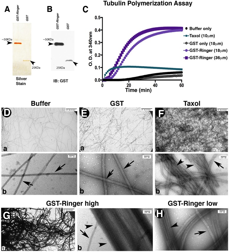
Ringer directly affects microtubule dynamics. (A,B) Representative silver-stain 12% gel (A) and anti-GST immunoblot (IB, B) showing purified proteins after the second round of purification for GST–Ringer (∼50 KDa, 2 μg) and GST alone (∼25 KDa, 1 μg) used for in vitro experiments. (C) In vitro tubulin polymerization assays, measured for optical densities (O.D.) at 340 nm and 37°C, showed purified GST–Ringer addition was sufficient to promote changes in microtubule polymerization rate and magnitude. (D–H) Representative images showing the ultrastructure of polymerization assay samples. Bottom panels in D–F are higher magnification images of those in the top panels. (D,E) Buffer and GST-only controls show unaided microtubule polymerization density. Single microtubules are marked with arrows (in Db,Eb,Fb,Gb,H). (F) Addition of Taxol increases microtubule density and promotes some bundling (arrowheads). (G,H) Addition of GST–Ringer to microtubules resulted in an increase of polymerization and had a microtubule-bundling effect (Ga,Gb,H, arrowheads). Scale bars: 10 μm (Da,Ea,Fa,Ga); 100 nm (Db,Eb,Fb,Gb,H).

