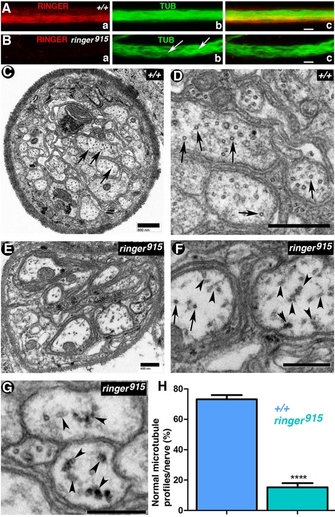
Fig. 7.

Lack of Ringer results in axonal microtubule defects. (A,B) Staining for α-Tubulin (TUB) and Ringer in +/+ and ringer915 third instar larval segmental nerves. Microtubules appear wavy and disorganized in mutants (Bb, arrows). Scale bars: 5 μm. (C–G) Representative transmission electron micrographs of +/+ (C,D) and ringer915-mutant segmental nerves (E–G). (C,D) In +/+ larval nerves, axonal microtubules (arrows) are normally observed as circular structures spread throughout the transverse face of the axon. Scale bars: 800 nm. (F,G) In ringer915 nerves, microtubule appearance (F,G, arrows) and distribution (F,G, arrowheads indicate instances of aberrant microtubule distribution) are compromised (F). F is a magnified image of an area of E. Scale bars: 400 nm. G shows a different example of microtubule accumulation in mutant nerves. Scale bar: 800 nm. (H) Quantification of the mean normal microtubule profiles per genotype as a percentage of the total per nerve shows that 84.74%±2.69 of +/+ (n=6) nerves exhibited a higher proportion of normal microtubules than the 26.84%±2.8 seen in ringer mutants (n=8) (****P<0.0001; Student's t-test).
