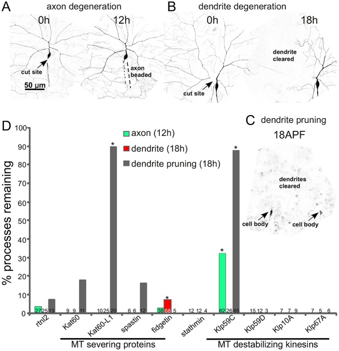
Fig. 1.

Identification of microtubule regulators required for degeneration and/or pruning. Class IV ddaC neurons were labeled with mCD8–GFP under the control of ppk-Gal4. (A) The assay for injury-induced axon degeneration is shown. 12 h after axon severing with a pulsed UV laser, axons were scored as beaded (normal degeneration) or continuous. (B) The assay for dendrite degeneration is shown. A ddaC dendrite was severed near its base with a pulsed UV laser at 0 h. The neuron was scored for complete clearance of the dendrite 18 h later. (C) Dendrite pruning was assayed 18 h after puparium formation (APF). An example image from a pupa is shown. Two adjacent cell bodies can be seen (arrows). Complete clearance of dendrites was scored as normal degeneration. (D) Tester animals (UAS-dicer2; ppk-Gal4, UAS-mCD8-GFP) were crossed with UAS-RNAi lines to target the microtubule regulators indicated. We used an RNAi line targeting rtnl2 as a control as it has never generated a phenotype in any assay we have performed. Kat60-L1 dendrite data was previously reported (Tao and Rolls, 2011), but is shown here for completeness. *P<0.05 compared to the appropriate rtnl2 control, all unmarked columns were not significantly different from controls (two-tailed Fisher's exact test). Numbers on each column are the number of animals tested for that condition.
