Fig. 4.
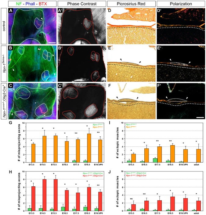
Phrenic axons misproject into the central tendon region and innervate ectopic muscles in both mutant embryos. Whole-mount diaphragm staining against neurofilaments and synaptophysin (green), actin (blue) and neuromuscular junctions (NMJs, red). At E16.5, the majority of misprojecting axons innervate ectopic muscles in the central tendon region of mutant embryos (B,C, dotted lines), and very scarcely also in wild-type embryos (A). Rare exceptions where an aberrantly projecting axon is not targeting ectopic musculature were observed (C, asterix). Phase contrast microscopy displays typically aligned myofiber orientation that is comparable to native costal muscles of the diaphragm (A′–C′). Picrosirius Red staining on frontal sections of E16.5 Npn-1Sema−/− (E) and Npn-1cond−/−;Olig2-Cre+ (F) mutant embryos shows that ectopic muscles (dotted lines) are lying on top of the central tendon region (dashed lines), whereas the majority of wild-type animals show an ectopic muscle-free central tendon region (D). Polarization microscopy further underpin the results, as they stretch on top of birefringent collagen fibers of the central tendon region (D′–F′). Arrowheads indicate the lack of collagenous extracellular matrix around ectopic muscles (E–F′). Scale bars: 200 µm (A–C′), 100 µm (D–F′). Quantification reveals significantly more axons that misproject into the central tendon region in Npn-1Sema−/− (G) and Npn-1cond−/−;Olig2-Cre+ (H) mutant embryos when compared to control embryos during the phase of diaphragm development. Interestingly, quantification of ectopic muscles in both mutant mouse lines reveals a slight, but not significant, increase of ectopic muscles until the beginning of muscle hypertrophy at E16.5, which then slightly decreased until adulthood (I,J). Data are represented as mean±s.e.m. (n≥3). *P≤0.05; **P≤0.01; ns, not significant (Mann–Whitney U-test).

