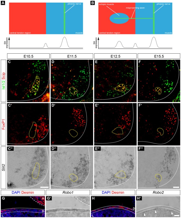
Fig. 5.
Slit2 and Robo1 are expressed in phrenic motor neurons and muscle progenitors, respectively, during diaphragm innervation. (A,B) Schematic view of hypothetical phrenic nerve and myoblast interaction during diaphragm development. In wild-type embryos, Slit is released from cells of the PPF at the border between central tendon region and premature muscle. In addition, Slit is also accumulated in the proximity of phrenic growth cones at the midline of newly formed costal muscles where it attracts resident muscle progenitors (A). In Npn-1Sema−/− and Npn-1cond−/−;Olig2-Cre+ mutant animals, axons that misproject into the central tendon region release Slit and attract some resident muscle progenitors, which later fuse to striated myofibers (B). Slit2 is strongly expressed in lateral motor column motor neurons (FoxP1+ cells, C′–F′,C″–F″) and a subpopulation of phrenic motor neurons (Scip+, Isl1+ and FoxP1−) between E10.5 and E15.5 (C,D,C″–F″). Although Robo1 (G′) is expressed within the desmin+ cells of the costal muscles (G), Robo2 (H′) is expressed by cells of the intermediate zone between the diaphragm and the liver (H, H′, arrow heads). H, heart; D, diaphragm; L, liver. Scale bars: 200 µm.

