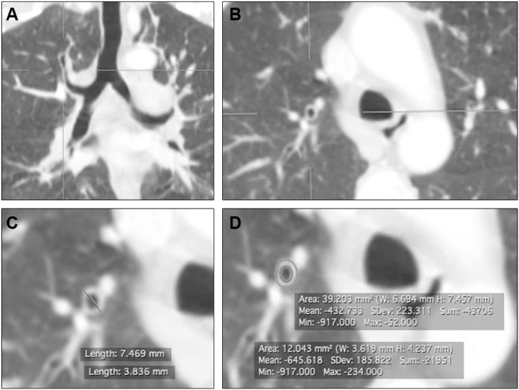
Figure 1.
Bronchial wall thickness measurement in high-resolution CT scan.
Notes: Coronal (A) and axial (B) cuts demonstrated the identification of apical segmental bronchus of right upper lobe bronchus (RB1). (C) Shows axial cut of chest CT showing circumferential measures of outer and inner areas of RB1 to calculate airway cross-sectional area (PWA%). (D) Shows axial cut of chest CT measuring the inner and outer diameters to calculate I/O ratio and T/D ratio ([inner diameter − outer diameter/2] to outer bronchus diameter).
Abbreviations: CT, computed tomography; I/O ratio, inner to outer diameter ratio; SD, standard deviation; W, width; H; height; Min, minimum; Max, maximum; T/D ratio, bronchial wall thickness to outer diameter ratio.

