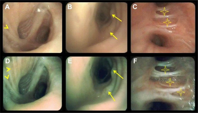
Figure 3.
HD WLB (A–C) and i-scan3 (D–F) showing various endobronchial mucosal changes.
Notes: A and D show mucosal striations (arrowheads), edema, and stoma; B and E show mucosal nodules (arrows); and C and F show mucosal thinning (stars).
Abbreviation: HD WLB, high-definition white light bronchoscopy.

