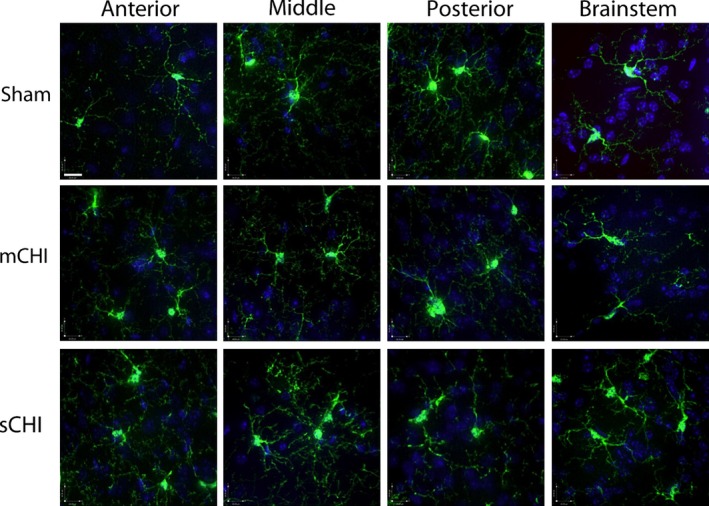
Figure 6.

Representative extended focus confocal images from the ipsilateral rostral (anterior) to caudal (posterior) regions of the cortices (clarified brain regions) and from the brainstem from sham, mild closed head injury (mCHI), and sCHI treated mice (Scale bar = 20.00 μm).
