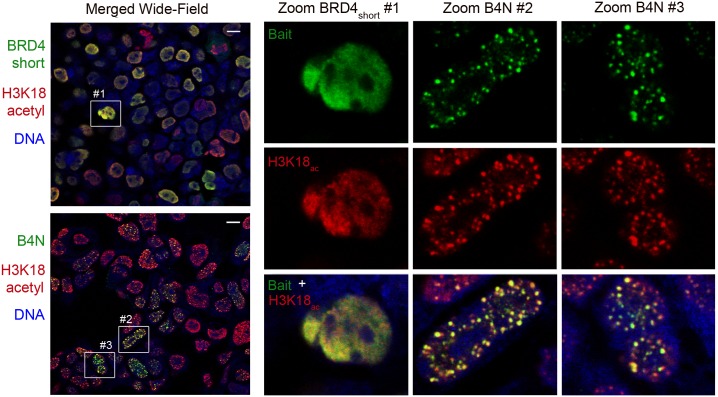
Fig 3. Formation of nuclear foci enriched in H3K18 acetylation by B4N and not BRD4short.
Confocal microscopy of 293-TREx cells with inducible HA-B4N and HA-BRD4short stained with anti-HA (green), which labels the bait protein, and anti-H3K18ac (red). Scale bar represents 10 micrometers. Zoomed-in images are provided to illustrate difference between H3K18ac granules in BRD4short cells and larger H3K18ac foci in B4N cells. See S5 Fig for non-confocal imaging and S6 Fig for separate green and red confocal layers of the same wide-field image of B4N-expressing cells.

