
Figure 3.
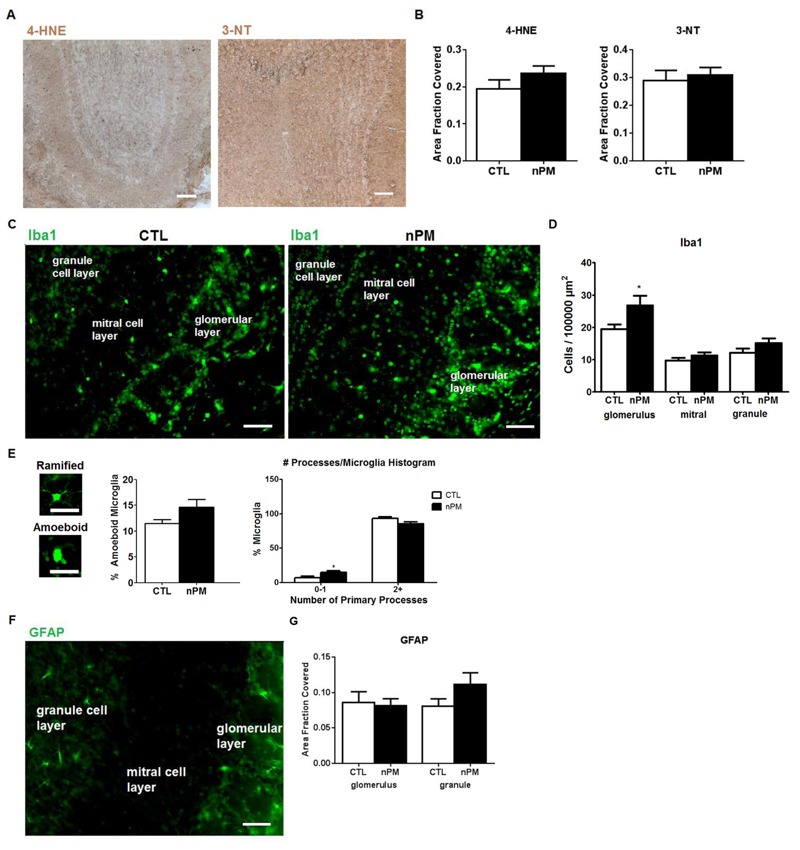
Acute in vivo nanoparticulate matter (nPM) exposure responses in olfactory bulb (OB): immunohistochemistry. CTL, control. (A) Immunostaining of 4-hydroxynonenal (4-HNE) and 3-nitrotyrosine (3-NT) in OB. (B) 4-HNE and 3-NT did not change significantly (n = 8). Scale bar = 100 μm. (C) Representative immunostaining of Iba1-positive macrophages. (D) nPM exposure increased the number of microglia in the OB glomerular layer by 30%, but no increase was observed in the mitral or granule cell layers. (*, p < 0.05; t-test). (E) Representative images of ramified versus amoeboid microglia. Scale bar = 25 μm. nPM doubled the percentage of microglia without multiple processes; the total percent of activated microglia did not change. [*, p < 0.05; two-way analysis of variance (ANOVA)]. (F) Immunostaining of astrocytes with glial fibrillary acidic protein (GFAP). (G) GFAP immunostained area by region was not altered by nPM exposure in any OB layer. Scale bar = 50 μm.