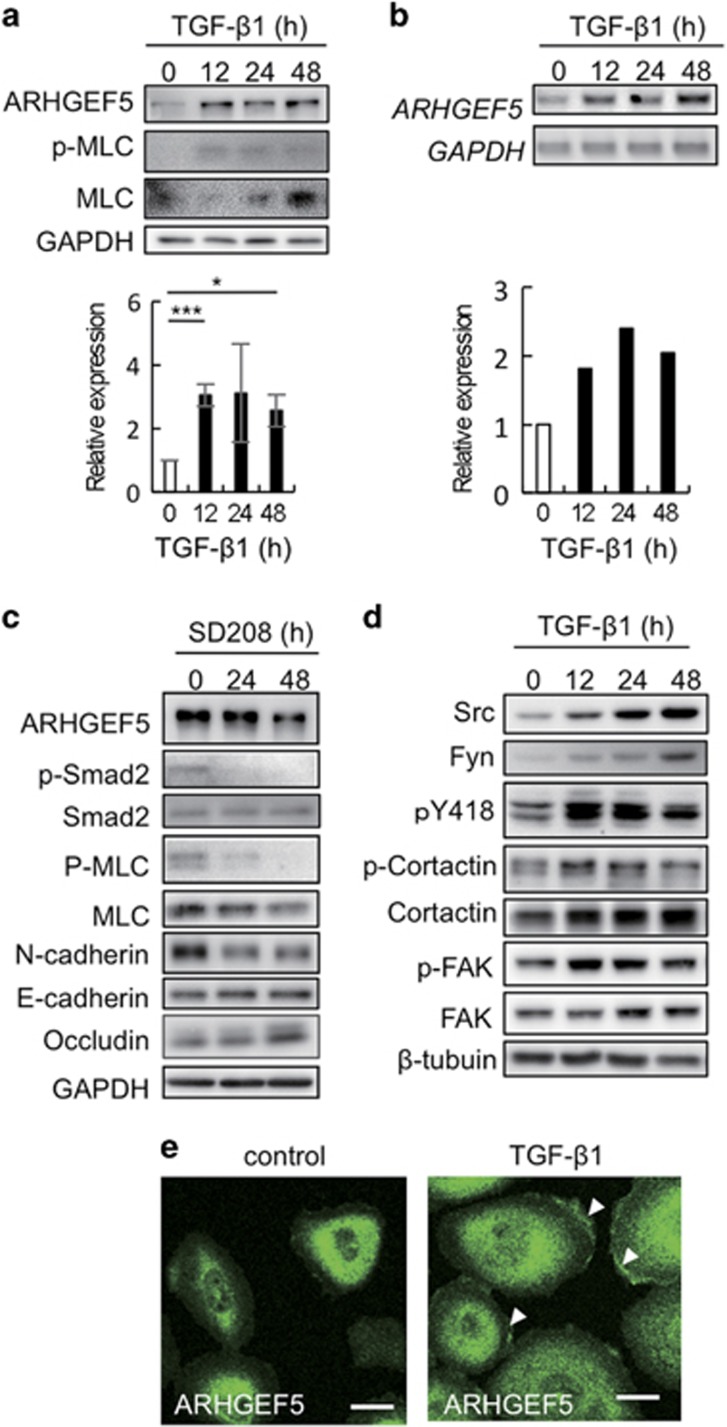
Figure 1.
ARHGEF5 and Src are upregulated during TGF-β-induced EMT in MCF10A cells. (a) Expression of ARHGEF5 and phospho-MLC in TGF-β1-treated MCF10A cells was analyzed by western blotting. Values represent the mean±s.d. (n=3, *P<0.05, ***P<0.001). (b) Expression of ARHGEF5 mRNA in TGF-β1-treated MCF10A cells was assessed by reverse transcription–polymerase chain reaction (RT–PCR). (c) MCF10A cells were treated with SD208 and the levels of the indicated proteins analyzed by western blotting. (d) Expression of the indicated proteins in TGF-β1-treated MCF10A cells was analyzed by western blotting. (e) MCF10A cells treated with or without TGF-β1 for 48 h were immunostained for ARHGEF5. White arrowheads indicate the ARHGEF5-positive areas in lamellipodia. Scale bar: 20 μm.

