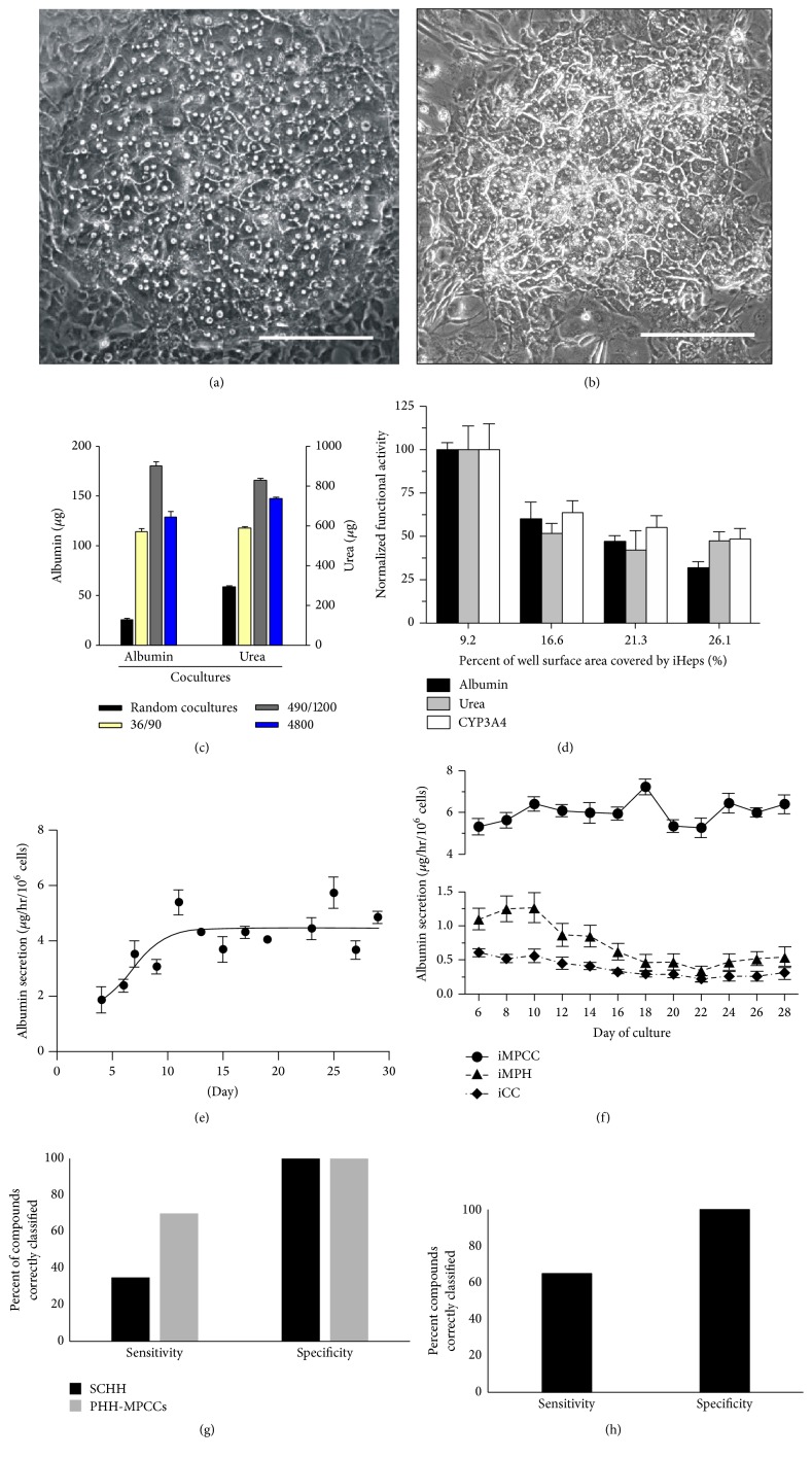
Figure 1.
Micropatterned cocultures (MPCCs) containing primary human hepatocytes (PHH) or induced pluripotent stem cell-derived hepatocyte-like cells (iPSC-HH) with supporting 3T3-J2 fibroblasts. Phase contrast images of PHH-MPCCs (panel (a)) and iPSC-HH-MPCCs (panel (b)) display similar hepatic morphology with polygonal shape, formation of bile canaliculi, and distinct nuclei/nucleoli. Scale bars on images represent ~250 µm. The architecture (island diameter, center-to-center spacing, percent of a well's surface area covered by hepatocytes) affects functions in both PHH-MPCCs (panel (c)) and iPSC-HH-MPCCs (panel (d)). In panel (c), cell numbers and ratios were kept constant while changing the diameter (first number) and center-to-center (second number) spacing of the PHH colonies [14]. In panel (d), total well surface area covered by the iPSC-HHs (also called iHeps) was modulated by changing the island diameter and spacing [116]. Albumin secretion levels can be maintained for at least ~1 month in PHH-MPCCs (panel (e)) [117] and iPSC-HH-MPCCs (circles in panel (f), triangles: micropatterned iPSC-HHs without 3T3-J2 fibroblasts, diamonds: iPSC-HH conventional confluent cultures) [116]. Compared to ECM sandwich-cultured primary human hepatocytes (SCHH), both PHH-MPCCs (panel (g)) and iPSC-HH-MPCCs (panel (h)) display higher sensitivity and similar specificity for drug toxicity screening when cultures were dosed for 6–9 days with a panel of 47 drugs [27]. Sensitivity for drug toxicity detection was 65% for iPSC-HH-MPCCs and 70% for PHH-MPCCs for the chosen drug set, while it was 35% for SCHHs. Permission was obtained from Nature Publishing group to reproduce panels (a), (c), and (e). Permission was obtained from John Wiley and Sons to reproduce panels (d) and (f).

