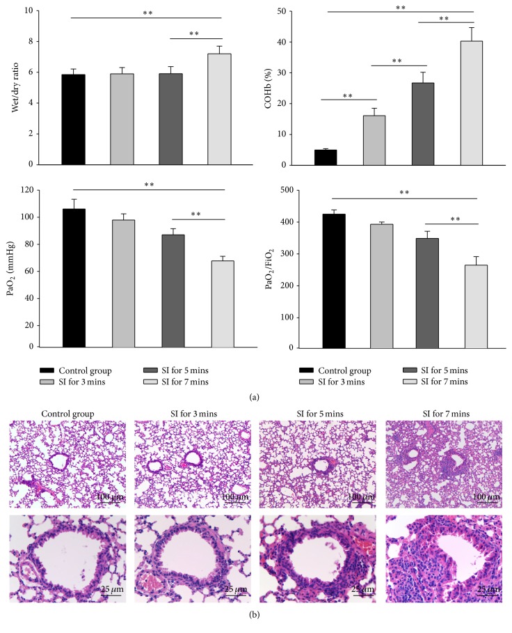
Figure 3.
Establishment of smoke inhalation NOD/SCID mouse model. NOD/SCID mice (n = 6) were subjected to 0, 3, 5, 7, and 9 min of smoke exposure. (a) Wet/dry (W/D) weight ratios, blood carboxyhemoglobin (COHb), PaO2, and PaO2/FiO2 were measured at the indicated time points. ∗∗ indicates P < 0.01 between the indicated groups. (b) Representative histological images of lung sections from mice with or without smoke inhalation injury (n = 6).

