
Figure 10.
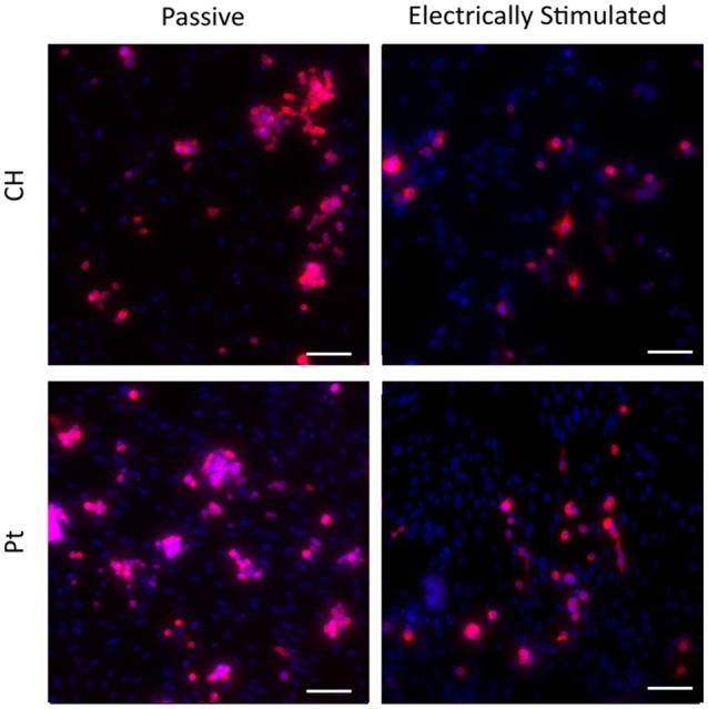
Magnified immunofluorescence images of PC12 and OEC co-cultures on passive and electrically stimulated Pt and CH substrates (200 × magnification). Nuclei of both cell types were stained with Hoechst 33,342 (blue) and PC12 only cell bodies and neurites stained with anti-βIII-tubulin (red). Scale bars = 150 μm.