
Figure 9.
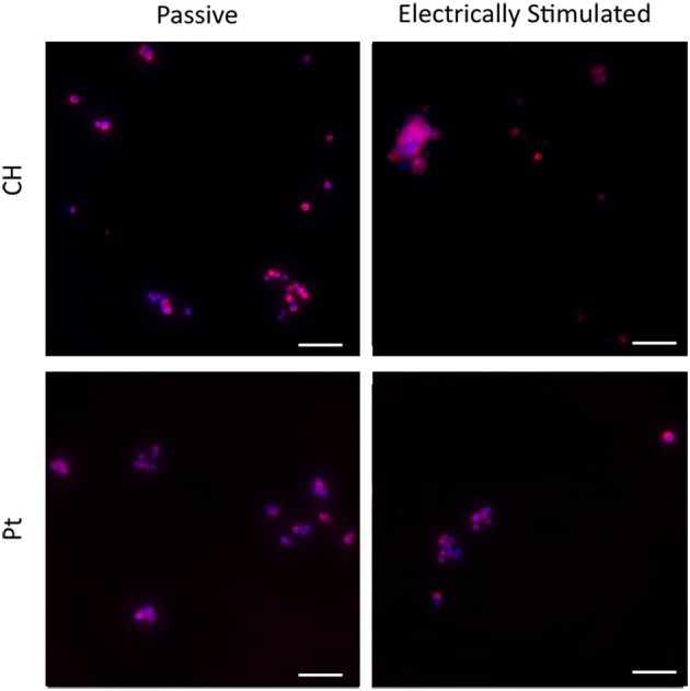
Magnified immunofluorescence images of PC12 cultures on passive and electrically stimulated Pt and CH substrates. Nuclei were stained with Hoechst 33,342 (blue) and PC12 cell bodies and neurites stained with anti-βIII-tubulin (red). Scale bars = 150 μm.