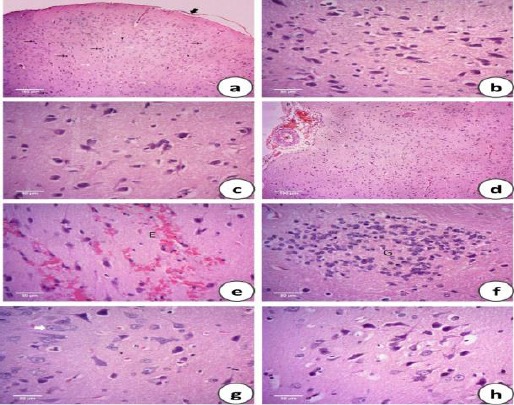
Figure 1.

Photomicrographs of the H&E stained brain sections of the control group (C) showing normal cerebral architecture with different sized intact basophilic neurons (black arrows) and randomly oriented axonal fibers (white arrows), normally distributed neuroglia cells (head arrows) and intact meningeal cell layers (thick arrow) (a). High magnification of the previous figure focused on the intact basophilic neurons (b). Propolis administered group (EEP) showing normal histological cerebral architecture (c). Heat stressed group (HS) showing meningeal cellular proliferation (P) with congested blood vessels (C) and mild submeningeal hemorrhage (H) (d), additional extravasated blood cells (E) were also observed between the neuronal tissues (e) with evidence of focal gliosis (G) detected by occasional aggregation of small rounded deeply basophilic neuroglia cells (f). Neuronal degeneration was observed and characterized by swollen rounded vacuolated neurons with pyknotic nuclei (white arrow) (g). The heat stressed group treated with propolis (EEP + HS) showing normal intact meninges and absence of both hemorrhage and gliosis with few to moderate numbers of neurons showing mild degenerative changes with occasional swelling and vacuolation (h)
