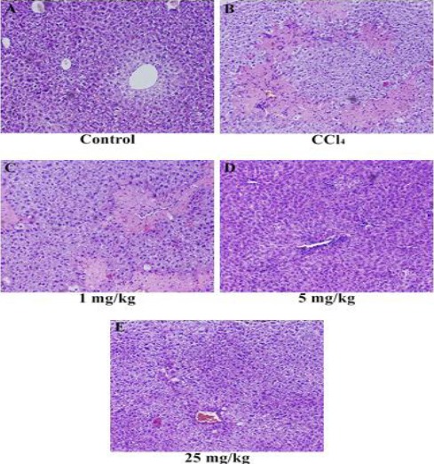
Figure 1.

Histopathology of liver tissues. (A) Liver section of normal control mice, showing normal architecture, (B) liver section of CCl4-treated mice, showing massive inflammatory cells and cellular necrosis, (C) liver section of mice treated with CCl4 and 1 mg/kg LCB, showing massive inflammatory cells and cellular necrosis, (D) liver section of mice treated with CCl4 and 5 mg/kg LCB, showing absence of necrosis and mild inflammatory cells, and (E) liver section of mice treated with CCl4 and 25 mg/kg LCB, showing the absence of inflammatory cells and absence of necrosis
