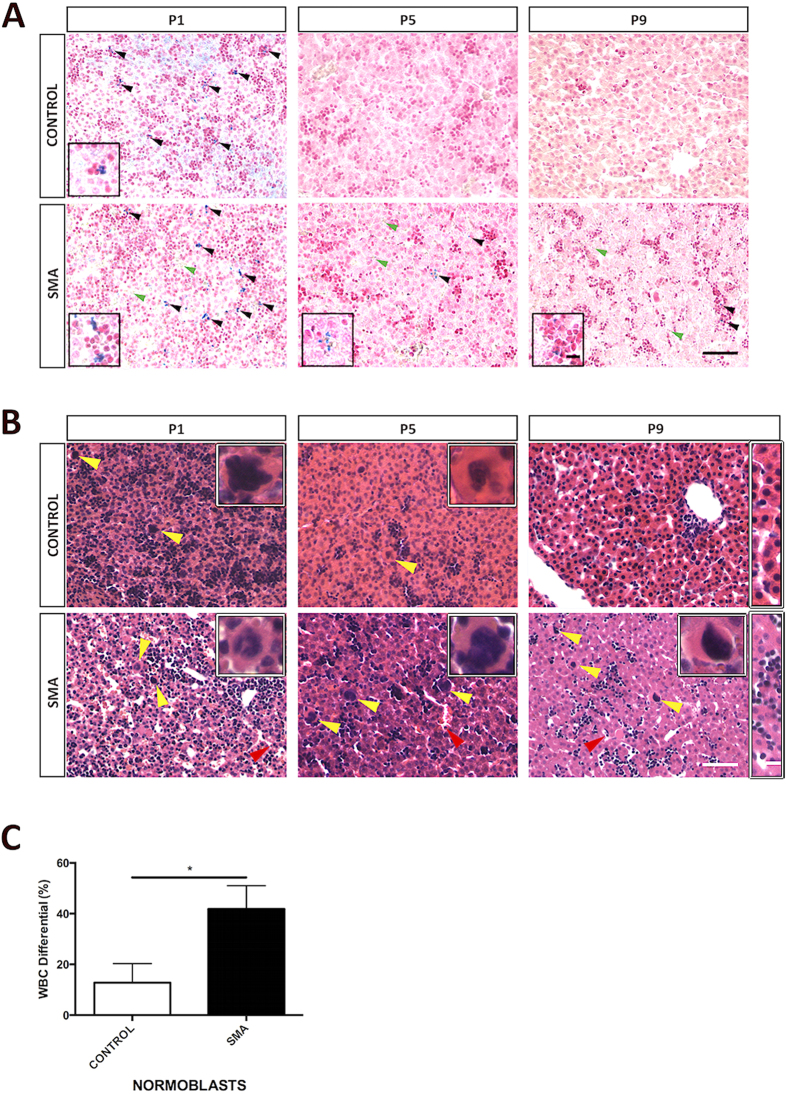
Figure 2. Persistent Erythropoietic Elements in SMA Liver Shows Developmental Failure.
(A) Representative light microscopy of Perl’s staining of livers at birth (P1) and postnatal days 5 (P5) and 9 (P9). Note the presence of iron deposits (black arrowheads) in later stages of development in SMA liver. P9 SMA liver appears to lack the organised hepatic plate structure as seen in control with predominant erythroblastic islands (dark red nuclei clusters). Green arrowheads point to RBCs outside main vessels. Magnified panels at bottom left show iron deposits (blue). Scale bar, 50 μm. Magnified panel scale bar, 10 μm. (B) Representative light microscopy of H&E-stained micrographs of livers. Red arrowheads show nucleated RBC within sinusoid. Yellow arrowheads and magnified panels at top right show megakaryocytes. P9 rectangular magnified panel shows nicely formed hepatic plate in control and lack thereof in SMA. Scale bar, 50 μm. Magnified panel scale bar, 10 μm. (C) White blood cell (WBC) differential showing the percentage of normoblasts in control and SMA blood samples obtained from P8 Taiwanese mice. p values were calculated using two-tailed Student’s t-test. Error bars, mean ± s.e.m. (n = 4 mice per group).

