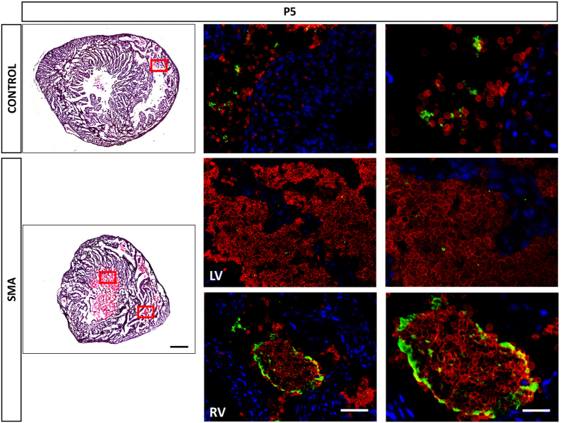
Figure 5. Platelets Aggregate into Circulating Clot-like Accumulations in SMA.
Representative micrographs of H&E and RBCs (Ly76+ DAPI− stain - red) and platelets (CD41+ stain - green) co-stained with DAPI (blue) of heart sections obtained at P5 from Taiwanese mice. Red squares show where Immunofluorescence images were taken. LV = Left Ventricle. RV = Right Ventricle. Note that the blood accumulation observed in SMA LV is not clotted unlike the one in RV. Scale bar for H&E, 500 μm. Scale bar for Immunofluorescence - left, 50 μm and - right, 10 μm.

