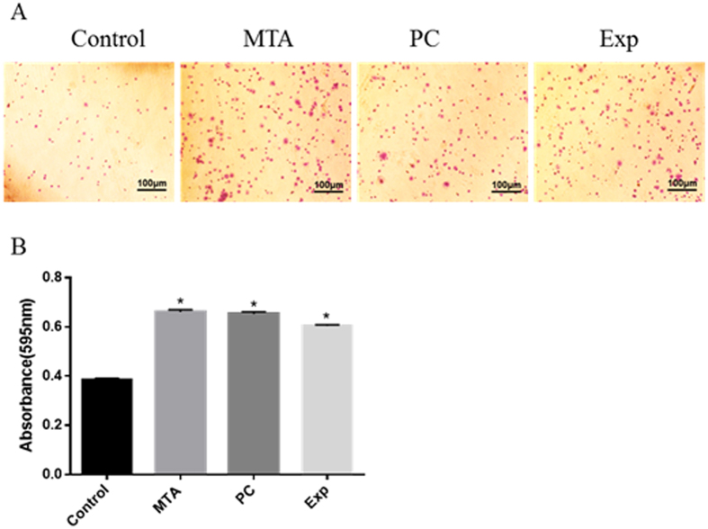
Figure 3. The effect of material extracts on adhesion ability in hDPSCs.
Cells were incubated with various material extracts for 1 h. Adherent cells were fixed and stained. The coloured solution was quantified at 595 nm on a microplate reader. (A) Representative diagrams for stained cells. (B) Quantification of the adherent cells. Data are presented as means ± standard deviation and measurements were performed in triplicate, with results summarised as the mean for each experiment. *P < 0.05 represents a significant change compared with the control.

