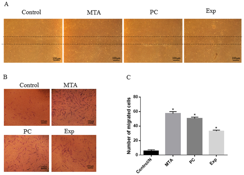
Figure 4. Effects of material extracts on migration in hDPSCs.
(A) Wound-healing assay. Cells were incubated with different material extracts for 24 h. Microphotographs of the scratches were obtained at 24-h post-wounding. (B) Cell migration assays were performed using a two-chamber Transwell system. Cells were treated with different material extracts for 24 h, and the migrated cells were fixed and stained. Representative photos of migrated hDPSCs were observed under a phase-contrast microscope. (C) Quantification of migrated cells. Data are presented as means ± standard deviation of three independent experiments. *P < 0.05 represents a significant change compared with the control.

