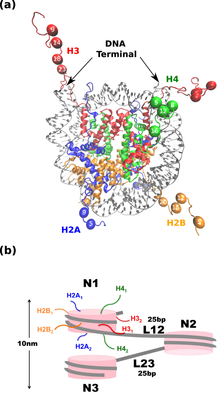
Figure 1. Mono- and tri-nucleosomes.

(a) X-ray crystallographic model of mono- nucleosome (PDB code 1KX5). Colors indicate different type of molecules: histone H3 is in red, histone H4 is in green, histone H2A is in blue, histone H2B is in orange, and DNA is in gray. Spheres represent lysine residues in N-terminal tails that can be acetylated and numbers in the spheres indicate residue numbers. Structural image is generated by VMD. (b) Cartoon image of tri-nucleosome. We call the first, the second, and the third nucleosomes as N1, N2, and N3, while the first and the second linker DNAs are called L12 and L23, respectively. Two copies of each histone are labeled by the subscript 1 and 2.
