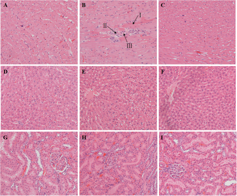
Figure 3. Histopathological examinations of heart, liver, kidney tissues in vehicle-, raw PR- and PRZA-treated rats, H&E staining, 100×.
(A) Heart tissue of control group: normal myocardial fibers in longitudinal section featuring central nuclei and syncytial arrangement of the fibers; (B) Heart tissue of raw PR group: myocardial fibers with loss of cross striations, not clearly visible nuclei, and inflammatory infiltration; (C) Heart tissue of PRZA group: the histopathological changes were milder than raw PR-treated group; (D) Liver tissue of control group; (E) Liver tissue of raw PR group; (F) Liver tissue of PRZA group; (G) Kidney tissue of control group; (H) Kidney tissue of raw PR group; (I) Kidney tissue of PRZA group. (D–I) Liver and kidney sections of both PR and PRZA groups showed no abnormalities. I. Myocardiocyte necrosis; II. Myocardiocyte rupture; III. Inflammatory infiltration.

