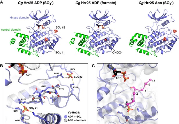
Figure EV2. binding sites in Hrr25.

- Aligned structures of Candida glabrata Hrr251–403 crystallized without nucleotide (Apo, left), or in the presence of ADP in crystallization buffer with formate (center) or sulfate (, right). / ions bound to sites 1 and 2 are shown as spheres. In the ADP (formate) structure, there is no ion bound to site 2, but a formate ion (CHOO−) is observed at site 1.
- Close‐up view of binding sites S1 and S2 in C. glabrata Hrr25 (blue), overlaid with the formate‐bound structure (white).
- View of the C. glabrata Hrr25 active site and at S1, with modeled substrate peptide from a Cdk2:cyclin A:peptide complex (PDB ID 1GY3) (Cook et al, 2002).
