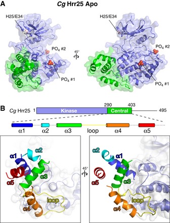
Figure 3. Structure of the Hrr25 central domain.

- Two views, roughly equivalent to the views in (A), of the Hrr25 central domain, colored as a rainbow according to the schematic at top. Dotted lines in the schematic indicate disordered loops (see Fig EV1A for sequence alignment of this domain).
