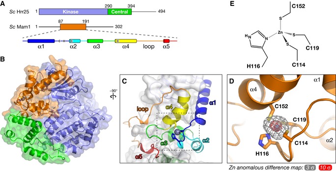
Figure 4. Structure of the Hrr25–Mam1 complex.

- Domain diagram of Saccharomyces cerevisiae Hrr25 (top) and Mam1 (bottom), with a schematic of Mam1 secondary structure. Zinc‐coordinating cysteine and histidine residues (Cys114, His116, Cys119, and Cys152) are shown in their approximate locations in the secondary structure (see Fig EV1B for Mam1 sequence alignment).
- Overall structure of the S. cerevisiae Hrr251–394 K38R:Mam187–191 complex, colored as in (A). View is equivalent to Fig 2B.
- Close‐up view of Mam187–191, colored as in the schematic in (A).
- Close‐up view of the variant zinc knuckle motif of Mam1, with zinc anomalous difference electron density shown at 3 σ (gray) and 10 σ (red).
- Geometry of zinc binding in Mam1. Zn‐S bonds and the Zn‐N bond were restrained to ˜2.3 Å and ˜2.0 Å, respectively, during refinement.
