
Figure EV1. Specificity of the CITK‐RNAi experiments.
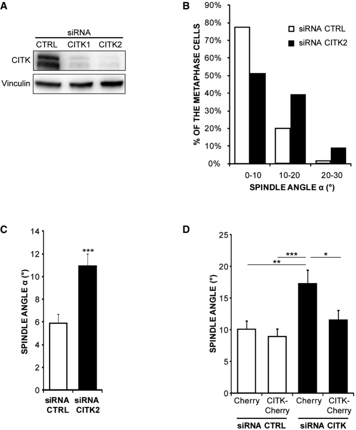
- Western blot analysis performed with the indicated antibodies on lysates of HeLa cells transfected with control or with two different CITK‐specific siRNAs (CITK1 and CITK2). The results of experiments shown in the main figures were obtained with the CITK1 sequence.
- Distribution of spindle angles (°) in HeLa cells depleted of CITK using the CITK2 sequence or transfected with control sequence. The values represent the angles between the axis crossing the two poles of metaphase spindles and the coverslip (n ≥ 150 cells in three independent experiments).
- Quantification of average and distribution of spindle angles (°) in HeLa cells depleted of CITK using the CITK2 (n ≥ 50 cells in three independent experiments).
- Quantification of angles average in HeLa cells transfected with either control or CITK1 siRNAs and cotransfected with expression plasmids expressing mCherry alone or fused to RNAi‐resistant (mouse) CITK wild‐type sequence (n ≥ 50 cells in three independent experiments).