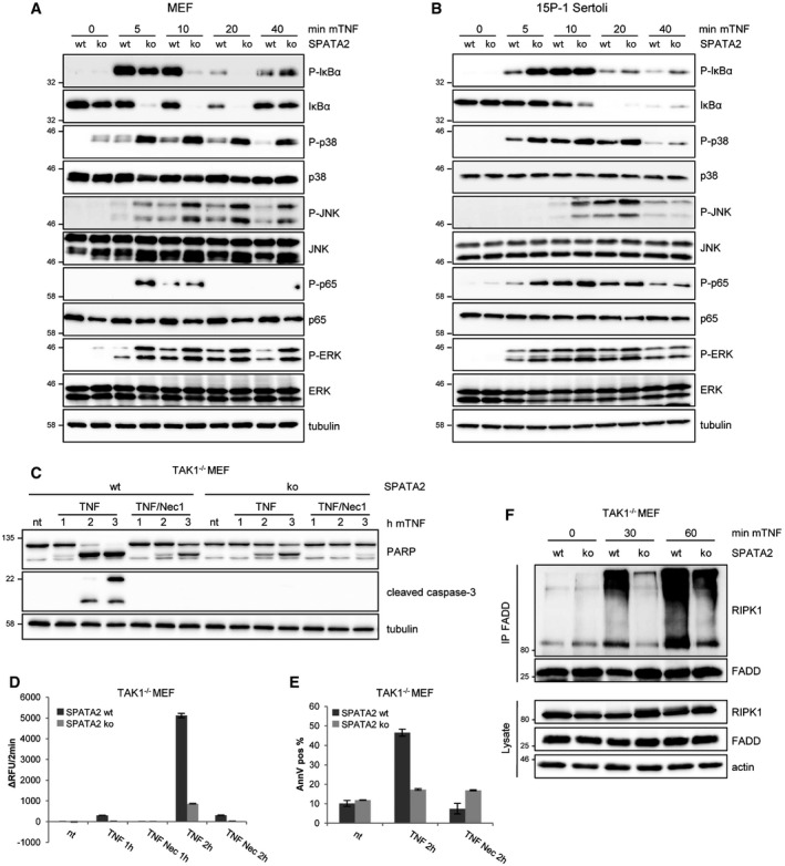
MEFs expressing CRISPR/Cas9 targeting luciferase (wt) or single clones generated from MEFs expressing CRISPR/Cas9 targeting the Spata2 gene (ko) were treated with mTNF (10 ng/ml) as indicated. The blots were probed with antibodies recognizing P‐IκBα, IκBα, P‐p38, p38, P‐JNK, JNK, P‐p65, p65, P‐ERK, ERK, and tubulin.
15P‐1 Sertoli cells expressing CRISPR/Cas9 targeting luciferase (as control) or single clones generated from 15P‐1 Sertoli cells expressing CRISPR/Cas9 targeting the Spata2 gene were treated with mTNF (10 ng/ml) as indicated. The blots were probed with antibodies recognizing P‐IκBα, IκBα, P‐p38, p38, P‐JNK, JNK, P‐p65, p65, and tubulin.
TAK1−/− MEFs were infected to express a CRISPR/Cas9 system targeting luciferase or the Spata2 gene, from which single clones were generated. The cells were treated with mTNF (10 ng/ml) or mTNF along with necrostatin‐1 (Nec‐1) for 1–3 h as indicated. The blot was sequentially probed with antibodies recognizing PARP, cleaved caspase‐3, and tubulin.
The same cells as used in (C) were treated as before with TNF (10 ng/ml) or TNF along with necrostatin‐1 (Nec‐1) for 1 or 2 h as indicated. Caspase activity was determined using the fluorogenic substrate DEVD‐AMC. A representative experiment is shown with error bars (representing SEM) referring to technical replicates.
Cells from the experiment as shown in (D) were analyzed for apoptosis by Annexin V staining after 2 h. A representative experiment is shown with error bars (representing SEM) referring to technical replicates.
Cells as described before were treated with mTNF (10 ng/ml) for the indicated time and lysates were subjected to immunoprecipitation with an anti‐FADD antibody. The immunoprecipitates were subjected to Western blotting and probed for RIPK1, FADD, and actin.