
Figure 2.
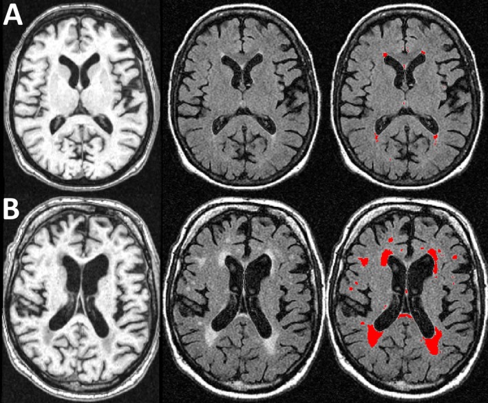
Magnetic resonance imaging (MRI) of persons with low (10th percentile) and high (90th percentile) burdens of white matter hyperintensities (WMH). Figure shows representative images of persons with low (10th percentile, row A) and high (90th percentile, row B) burdens of WMH. The first column (L) shows T1‐weighted MPRAGE axial images, the second (middle) shows T2‐weighted FLAIR images, and the third (R) shows the segmented WMH lesions in red (3rd) column.