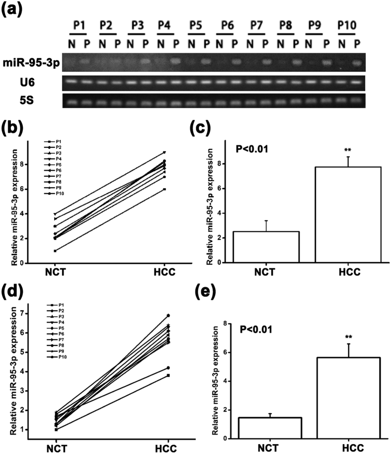
Figure 8. The expression level of miR-95-3p is higher in HCC tissues than in normal adjacent non-cancerous tissues.
(a) Semi-quantitative RT-PCR analysis showed a higher expression level of miR-95-3p in HCC tissue samples than in normal adjacent tissue samples. (b) PCR bands in (a) were quantified and plotted. The ratio of the expression level of miR-95-3p over U6 RNA in HCC tissue sample was higher than that in the normal adjacent non-cancerous tissue sample in all 10 groups. (c) The relative miR-95-3p expression level over U6 RNA in human HCC tissues was significantly higher than that in normal adjacent non-cancerous tissues. (d) PCR bands in (a) were quantified and plotted. The ratio of the expression level of miR-95-3p over 5S RNA in HCC tissue sample was higher than that in the normal adjacent non-cancerous tissue sample in all 10 groups. (e) The relative miR-95-3p expression level over 5S RNA in human HCC tissues was significantly higher than that in normal adjacent non-cancerous tissues.

