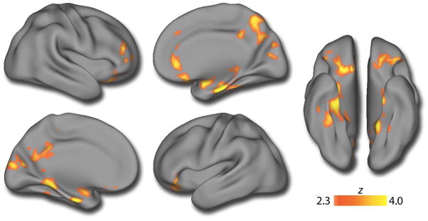
Figure 2.
Multifocal gray matter volume reduction in psychosis-spectrum youth. A voxelwise between-group analysis of gray matter volume reveals that PS youth have diminished GM volume across multiple brain regions, including the precuneus, posterior cingulate, bilateral medial temporal lobes, frontal pole, and orbitofrontal cortex. Image thresholded at z>2.3, corrected p<0.01; minimum cluster size: k>255 voxels. See eTable 1 for further details.

