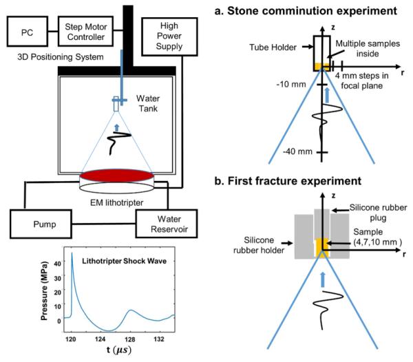
Figure 1.

Diagram of the experimental setup: (a) positions of the tube holder during stone comminution experiments using different size groups of approximately matched total mass (b) the stone holder and placement of individual stone of different sizes to determine the number of shocks required to produce the first fracture.
