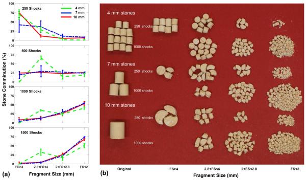
Figure 6.
(a) Variations of fragment size (FS) distributions in three different stone size groups after treatment in water at the lithotripter focus. (b) Representative photos of original stones of different sizes (left column) and corresponding fragment distribution in different size ranges after 250 shocks and 1000 shocks.

