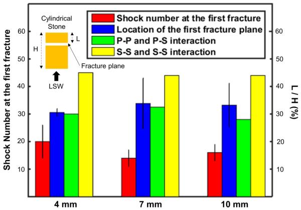
Figure 7.
The number of shocks delivered to produce the first fracture in individual 4-, 7-, 10- mm cylindrical stones (red bar), locations of the first fracture plane (blue bar), locations of P-P wave and P-S wave interaction (green bar), and location of S-S and S-S wave interaction (yellow bar). Note, P-S: Shear waves generated by the reflection of incident longitudinal waves; P-P: Longitudinal waves generated by the reflection of incident longitudinal waves; S-S: Shear waves generated by the reflection of shear waves.

