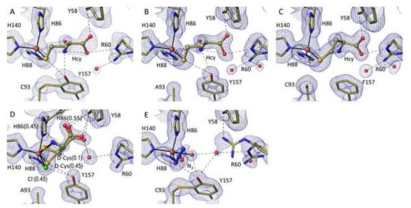
Figure 6.
High resolution views of the homocysteine (Hcy), D-Cys, and azide complexes of wild-type and/or mutant CDOs. 2Fo-Fc electron density and models and interactions are as in Figure 2. (A) Hcy soak of wild-type CDO at pH 8.0 (PDB code 4XF4). (B) Hcy soak of C93A at pH 8.0 (PDB code 4XF9). (C) Hcy soak of Y157F at pH 8.0 (PDB code 4XFA). (D) D-Cys soak of C93A at pH 8.0 (PDB code 5I0R). The orientation is rotated slightly relative to the other figures to better show the His86 alternative conformation. The bound D-Cys α-amino group would collide with His86 in its iron ligating position (being ~1.6 Å from His86-Nε2). (E) Azide soak of wild-type CDO at pH 6.2 (PDB code 4PJY).

