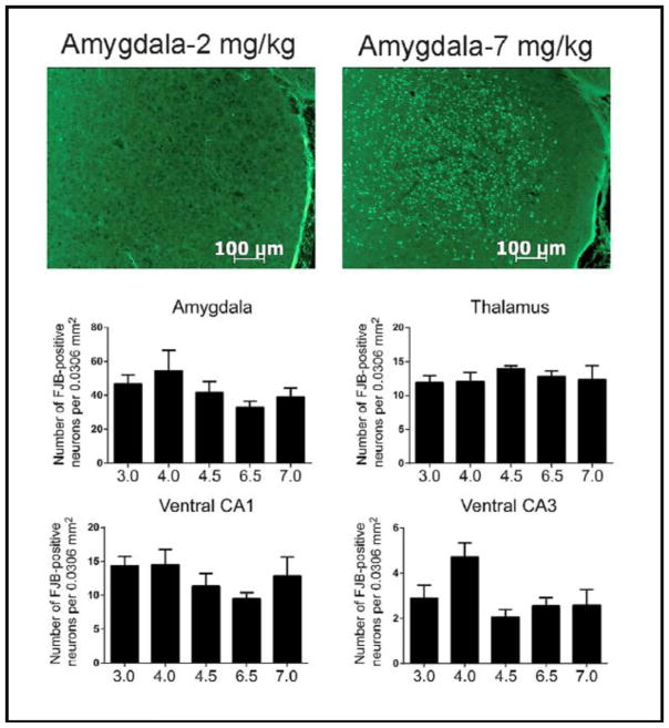
Fig. 5.
Top Panels - Representative sections stained with Flouro-Jade B from the amygdalae of animals given 2 mg/kg (left panel) or 7 mg/kg (right panel) DFP. Middle and Lower Panels - Summary data showing the numbers of FJB stained cells in representative brain regions from animals given 3–7 mg/kg DFP. (N’s= 3.0 mg/kg, 10; 4.0 mg/kg, 9; 4.5 mg/kg, 10; 6.5 mg/kg, 9; 7.0 mg/kg, 10)

