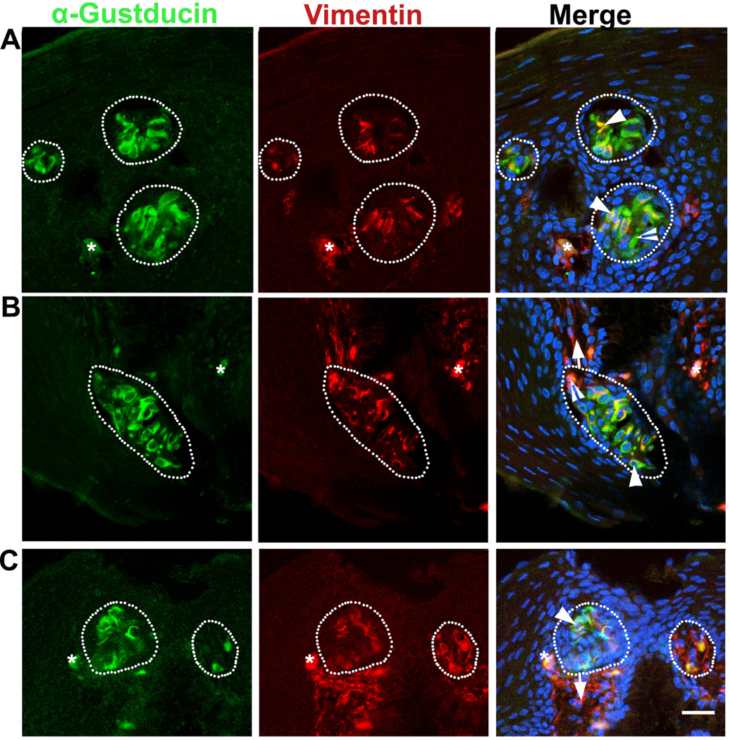
Fig. 2.
Distribution of α-Gustducin (green) and Vimentin (red) immunosignals in the oral tissue of a P0 chicken: base of the oral cavity (A), palate (B), and posterior region of the tongue (C). White dots outline the specified cell clusters, presumably taste buds. Arrows point to the connective tissue. Arrowheads in A–C point to some of the cells double labeled with α-Gustducin and Vimentin. Open arrowheads in A–B point to singly labeled α-Gustducin+ or Vimentin+ cells. Asterisks in A and C mark cells with nonspecific labeling. Scale bar: 20 µm for all images (single plane laser-scanning confocal).

