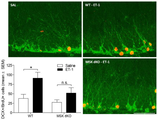
Figure 4. Ischemia-induced neurogenesis.
Post-ischemic neurogenesis was assessed 4 weeks following ET-1 or saline infusion. Newborn neurons were labeled immunohistochemically for doublecortin (green) and Brdu (red). Data are presented as the total number of BrdU-positive cells that are co-labeled with doublecortin. The number of newborn neurons is significantly increased in WT but not MSK dKO mice (F3,21 = 4.913, p<0.05). Scale bar = 50μm. Data are presented as mean ± SEM. An asterisk (*) denotes a statistically significant difference between groups, n.s. denotes that the group comparison is not significant.

