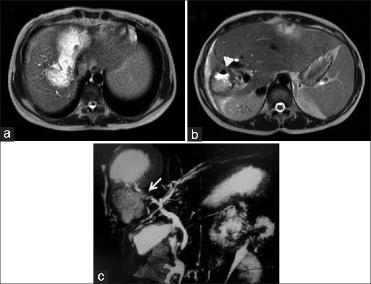
Figure 11.

Magnetic resonance imaging axial T2-weighted (a and b) and magnetic resonance cholangiopancreatography (c) images show communication between ruptured hydatid cyst and biliary radicle (arrow) with air-foci within the cyst (arrow head) and biliary radicles. Right lobe of liver atrophy is noted
